Related News


CZI grant awarded to UCL and ESRF project for the development of HiP-CT imaging
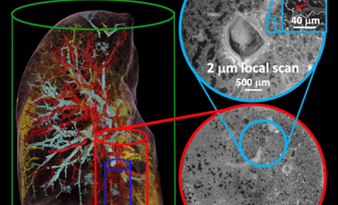
The 54-year-old male’s right upper lung lobe was first scanned at 25 micron voxel resolution (green cylinder, rendered to show the two vascular systems and occluded vessels). Scientists then zoom in at 6 (red circle) then 2 micron voxels (blue circle), giving 100X more resolution than clinical CT. Cellular structure is resolved, including individual red blood cells (red arrows). (Credit: P.Tafforeau/ESRF).