Automatic analysis of cryo-electron tomography
Introduction
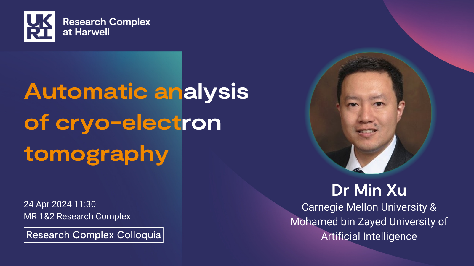
Seminar Abstract: The cell is the basic structural and functional unit of all living organisms. Understanding how cells function is fundamental to life science. Macromolecules are nano-machines inside cells that govern the cellular processes. To fully understand such processes, it is necessary to know the native structures and spatial organizations of macromolecules inside single cells, and their interactions with other subcellular components. Such information has been extremely difficult to obtain due to a lack of suitable data acquisition techniques.
The recent revolution of Cryo-electron tomography (cryo-ET) 3D imaging technology has made collecting such information possible. Cryo-ET captures a 3D image of a single cell's subcellular structures at sub-molecular resolution and in a near-native state. It provides unprecedented opportunities for systematically studying the native spatial organization of subcellular structures, especially macromolecules. However, cryo-ET has a high degree of structural complexity and imaging limits, such as high structural diversity and crowding, low signal-to-noise ratio, and missing values. These have made the automated systematic analysis of such images extremely difficult.
Since 2008, we have been developing image analysis methods to address this challenge. In particular, we focus on systematic recognition and recovery of the structures of large numbers (millions) of macromolecules captured by cryo-ET, without relying on external structural knowledge. To do so, we have developed different cryo-ET image registration, classification, segmentation restoration techniques. Our effort is a key step for systematic analysis of macromolecules' structures and spatial organizations inside single cells captured by cryo-ET.


