

Ductile cast irons (DCIs) are becoming increasingly important for industries working in renewable energy and transportation. They are stronger, tougher and less expensive than gray cast iron, which is commonly used for automotive brake systems as it transfers heat well and has excellent machinability qualities. Whilst strong, gray iron can be brittle, whereas DCI is more resilient when undergoing stresses related to thermo-cycling or impact, so can be used for steering, suspension and gears. DCIs are now being used for several components in wind turbines, with ever increasing demands being placed on their mechanical and fatigue properties.
With DCIs growing in the use of applications, the Materials Design and Manufacturing Group at Research Complex at Harwell, led by Professor Peter Lee at UCL, have been examining what dictates the mechanical performance of DCIs. Synchrotron X-ray light sources have evolved dramatically over the last two decades. With the incorporation of fast acquisition systems, it is now possible to capture a vast amount of rich microstructural information to unravel various underlying mechanisms.
In situ 3D tomography has been used for solidification investigations in low melting point alloys for more than a decade. The development of advanced high temperature environmental cells and high X-ray contrast alloy systems has led to the creation of a toolset for manufacturers to investigate patterns and any potential problems. In order to build robust global microstructure prediction tools, it is necessary to understand normal and abnormal growth of graphite nodules in DCIs, as well as the effect of thermal fluctuations.
A joint project with Dr. Mohammed Azeem at Leicester University and a team at the Danish Technical University (DTU) was formed, utilising the capabilities developed in the RCaH. In this investigation led by Dr. Tim Wigger at Research Complex, the regular growth of nodules was captured with a temporal resolution ten times faster than previous experiments the group had worked on. The experiment was performed at the I12-JEEP beamline of Diamond Light Source. The beamline provides high energy and photon flux for high-speed tomography. A ductile cast iron alloy was used in the experiments, containing 1.94 ± 0.02 wt.% Si for stabilisation of the graphite and 0.075 ± 0.005 wt.% Mg to enable the formation of graphite nodules.
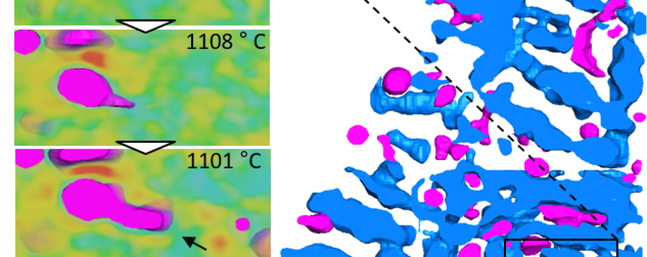
The degeneration of nodules was observed by the team to increase with re-melting cycles, which is attributed to Mg-loss. Austenite shells around graphite nodules seemed to form with varying thickness. The tomography data indicates that there are thinner regions (or open channels) through which carbon can diffuse easily. Graphite protrusions formed and grew through these higher carbon concentration channels which led to nodule degeneration. Large pre-existing nodules can break free and develop star-shapes whilst floating up into hotter regions.
Dr. Tito Andriollo at DTU developed a 2D axisymmetric finite element model based on carbon diffusion, which indicates that the shell with thin sections can deform and open as the nodule grows. This supports the team’s experimental observations and theories of liquation cracking of the austenite shell during solidification. These results provide valuable insights into the solidification kinetics of cast irons, supporting the design of advanced alloys.
T. Wigger, T. Andriollo, C. Xu, S.J. Clark, Z. Gong, R.C. Atwood, J.H. Hattel, N.S. Tiedje, P.D. Lee and M.A. Azeem, In situ synchrotron investigation of degenerate graphite nodule evolution in ductile cast iron, Acta Materialia, Volume 221, December 2021, 117367.
Image from Graphical Abstract: Graphite features embedded in austenite dendrites. Formation of degenerate nodule and evolution of carbon concentration field during solidification.